Honey has been used for over 5000 years. Why has such a product been utilized for so long? I would suggest that it is due to its brain health and well-being properties.
Mineral interaction with Pi electrons of aromatic rings
Research from the past two decades has explored honey as an enigmatic gel that has gastroprotective, hepatoprotective, reproductive, hypoglycemic, antioxidant, antihypertensive, antibacterial, antifungal, anti-inflammatory, immunomodulatory, wound healing, cardio-protective and antitumor effects [6, 26, 36, 37]. Unfortunately, research on the nootropic and neuropharmacological effects of honey is scarce.
Nevertheless, the belief that honey is a memory-boosting food supplement is actually ethnotraditional as well as ancient in nature. For instance, honey is reported to be an important component of Brahma rasayan, an Ayurvedic formulation that is prescribed to extend the lifespan and improve memory, intellect, concentration, and physical strength [38].
One established nootropic property about honey is that it assists the building and development of the entire central nervous system, particularly among newborn babies and preschool age children, which leads to the improvement of memory and growth, a reduction of anxiety, and the enhancement of intellectual performance later in life [26].
Additionally, the human brain is known to undergo postnatal development with the obvious maturation and reorganization of several structures, such as the hippocampus and cerebral cortex. It has been reported that this postnatal development occurs through neurogenesis, which occurs predominantly during childhood, and this development can also extend into adolescence and even through adulthood [39]. Empirical, but striking, evidence supporting this concept was provided by an experiment that was conducted on postmenopausal women; those who received honey showed improvements in their immediate memory but not in immediate memory after interference or in delayed recall [40]. In another study, the normal diet of two-month-old rats was supplemented with honey, and their brain function was assessed over a one-year period. Honey-fed rats showed significantly less anxiety and better spatial memory throughout all stages compared with the control group of rats. More importantly, the spatial memory of honey-fed rats, as assessed by object recognition tasks, was significantly greater during later months (i.e., 9 and 12) [41].
Restoring brain health and mental well-being.
OH BEE HAVE Manuka honey spray produces high energy short lived radicals that have sufficient energy to enable the restoration of brain health and wellbeing. The brain health benefits associated with honey have predominately been attributed to the phenolics present in the honey.
Examples include:
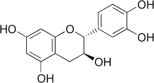
Apigenin is a common flavonoid that is frequently identified in honey. In addition to its radical scavenging activity, apigenin protects neurons against oxygen-glucose deprivation/reperfusion-induced injury in cultured primary hippocampal neurons by improving sodium/potassium-ATPase (Na+/K+-ATPase) activities [58]. Apigenin also inhibits the kainic acid-induced excitotoxicity of hippocampal cells in a dose-dependent manner by quenching reactive oxygen species and by inhibiting the depletion of reduced glutathione (GSH) levels [59]. Apigenin suppresses the interferon gamma (IFN-𝛾)-induced expression of CD40, whereas the signaling of CD40 is critically involved in microglia-related immune responses in the brain. Rezai-Zadeh et al. suggested that apigenin may have neuroprotective and disease-modifying properties in several types of neurodegenerative disorders [60]. Moreover, apigenin stimulates the adult neurogenesis that underlies learning and memory [39]. Apigenin is a flavonoid belonging to the flavone structural class and chemically known as 4′,5,7,-trihydroxyflavone, with molecular formula C15H10O5. Apigenin has a low molecular weight (MW 270.24), structurally forming yellow needles in pure form.
The neuroinflammatory suppression activity of caffeic acid can be inferred from the observation that caffeic acid reverses the aluminum-induced overexpression of 5- lipoxygenase (5-LOX) in brain tissues [62]. Caffeic acid, another important antioxidant, is a type of phenolic acid that is present in honey, as well as in coffee, fruits and vegetables. An in vitro study has demonstrated the neuroprotective effects of caffeic acid on neuronal cells [61]. Caffeic acid also prevents the aluminum-induced damage of the cerebrum that is associated with neuronal death in the hippocampus and with learning and memory deficits [62]. In vitro treatment with caffeic acid at several different concentrations has been reported to increase the acetylcholinesterase activity in the cerebral cortex, cerebellum, and hypothalamus. A similar scenario is also observed in the cerebellum, hippocampus, hypothalamus, and pons when caffeic acid is administered in vivo. All of these findings strongly support the proposition that caffeic acid improves memory by interfering with cholinergic signaling, in addition to its neuroprotective effects [63].
Catechin is a flavonoid that contributes to the antioxidant activities of honey. Several studies have repeatedly demonstrated the neuroprotective effects of catechin on neuronal death in a wide array of cellular and animal models of neurological diseases [64, 65]. Although catechin possesses potent iron-chelating, radical-scavenging, and anti-inflammatory activities, current studies have indicated that the modulation of signal transduction pathways, cell survival, or death genes and mitochondrial function significantly contribute to the induction of cell viability [66]. For instance, according to Unno et al., the daily consumption of green tea, which contains high levels of catechin, can delay the memory regression Evidence-Based Complementary and Alternative Medicine 5 that is associated with age-related brain atrophy and cognitive dysfunction [67]. Animal studies have indicated that the long-term administration of green tea may prevent agerelated learning and memory decline by modulating the transcription factor cAMP-response element binding protein (CREB) and by upregulating synaptic plasticity-related proteins in the hippocampus [68, 69]. Similar memoryameliorating effects were also shown in the context of neurodegenerative diseases, such as PD, AD, and multiple sclerosis [64].
Chlorogenic acid is a derivative of caffeic acid and is another common phenolic acid that is found in honey. A dose-dependent protective effect of chlorogenic acid against apoptosis was observed in pheochromocytoma-12 (PC12) cell lines that were exposed to methyl mercury-induced apoptotic damage. The protective activity of chlorogenic acid was associated with a reduction in the generation of reactive oxygen species (ROS) and the attenuation of apoptosis by the activation of caspase-3 [70]. In a study by Kwon et al. [71], the neuroprotective effects of chlorogenic acid on scopolamine induced learning and memory impairment were using several behavioral tests, such as the Y-maze, avoidance, and Morris water maze tests.
Chlorogenic acid inhibits the synthesis and release of inflammatory mediators, such as tumor necrosis alpha and nitric oxide (NO), thus contributing to anti-inflammatory and analgesic activities against carrageenan-induced inflammation [72]. Therefore, the chlorogenic acid in honey might have the capacity to attenuate neuroinflammation.
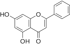
Chrysin (5,7-dihydroxyflavone) is another important flavonoid antioxidant that is present in honey. A behavioral experimental model revealed that chrysin is an anxiolytic that acts as a central receptor for benzodiazepine in instances where anxiety was reported to hamper cognitive function and learning capacity [73]. A study conducted by He et al. [74] showed that the therapeutic potential of chrysin in neurodegeneration-associated dementia resulted from cerebral hypoperfusion. The effects of chrysin were further investigated in a rat model of cognitive deficits and brain damage generated by the permanent occlusion of the bilateral common carotid arteries [74]. Such surgically induced hypoperfusion leads to a significant increase in the escape latency in the Morris water maze, with biochemical features of neural damage, such as increases in glial fibrillary acidic protein expression and apoptosis. Interestingly, chronic treatment with chrysin significantly alleviated neuronal damage and spatial memory deficits, with a reduction in lipid peroxidation and glutathione peroxidase activity but a decrease in SOD activity [74], indicating the neuroprotective role of honey.
Ferulic acid could also alleviate learning and memory deficits through the concomitant inhibition of acetylcholinesterase activity and the augmentation of SOD activity while lowering the concentration of glutamic acid and malondialdehyde in the hippocampus of rats.These results suggested that the antioxidant activities of the honey may contribute to the improvement of the cholinergic system in the brain or to the inhibition of nerve injury by excitatory amino acids [82]. Ferulic acid may be useful for preventing trimethyltin-induced cognitive dysfunction as well as for boosting the activation of choline acetyltransferase (ChAT) in dementia [83].
Gallic Acid. Gallic acid prevents the apoptotic death of cortical neurons in vitro by inhibiting amyloid beta (25–35)-induced glutamate release and the generation of ROS [84]. Gallic acid possesses an antianxiolytic activity, which provided the primary evidence in support of the memory-ameliorating effect of gallic acid because anxiety is associated with memory disturbance [85]. The memory-ameliorating effects of gallic acid were further confirmed by Al Mansouri et al. [86], who revealed its neuroprotective effect on 6-hydroxydopamine-induced and cerebral oxidative stress-induced memory deficits. Gallic acid improved memory concomitant with increases in the total thiol pool and glutathione peroxide activity and decreased lipid peroxidation in the hippocampus and striatum [87]. However, we cannot claim that these biochemical findings are entirely responsible for the improvements in memory.
Kaempferol is a plant flavonoid that is also frequently found in honey. The toxicity of 1-methyl-4-phenyl-1,2,3,6- tetrahydropyridine (MPTP), which is a neurotoxin, leads to behavioral deficits, a depletion of dopamine, reductions in SOD and glutathione peroxidase activities, and an elevation of lipid peroxidation in the substantia nigra. The administration of kaempferol has been reported to reverse all of these behavioral and biochemical alterations and to prevent the loss of TH-positive neurons that is induced by MPTP (1-methyl-4-phenyl-1,2,3,6-tetrahydropyridine) [88].
In another study, kaempferol demonstrated the ability to protect primary neurons from rotenone-induced apoptotic challenge. Specifically, kaempferol-ameliorated antioxidant defenses and antiapoptotic effects involve the enhancement of mitochondrial turnover, which is mediated by autophagy [89]. Furthermore, kaempferol may be an optimal treatment for improving cognitive function due to its positive effects on depression, mood, and cognitive functions [90].
Luteolin is a flavonoid from the flavone class that has been reported to be found in honey. As is the case for most flavonoids, luteolin has antioxidant, anti-inflammatory, and antitumor properties [91]. Luteolin also has neuroprotective effects against microglia-induced neuronal cell death. The consumption of luteolin has been found to improve the spatial working memory of aged rats by mitigating microglia-associated inflammation in the hippocampus [92]. The impairment of learning acquisition induced by cholinergic neurotoxins and muscarinic and nicotinic receptor antagonists were reported to be attenuated by luteolin. This phenomenon, however, was not observed for dopaminergic neurotoxin- and serotonergic neurotoxin-induced memory impairments, thus confirming the involvement of the central cholinergic system in the memory-restoring function of luteolin [93].
Interestingly, Tsai et al. showed that the augmenting effect of luteolin on Mn-SOD and (Cu/Zn)-SOD activity as well as on the GSH levels in the cortex and hippocampus was associated with the amelioration of amyloid beta (1– 40)-induced oxidative stress and cognitive deficits [94].
This last observation is very interesting when one understands the links between dementia, Alzheimer's disease and Downs Syndrome.
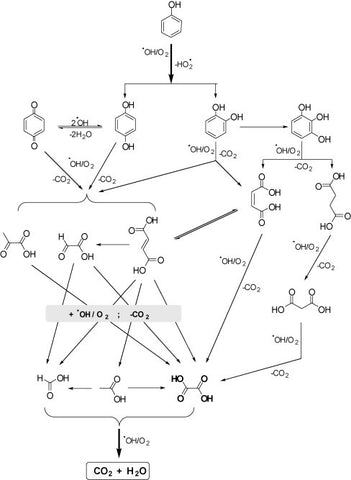
As with all adults, advancing age also increases the chances a person with Down syndrome will develop Alzheimer's disease. According to the National Down Syndrome Society, about 30% of people with Down syndrome who are in their 50s have Alzheimer's dementia. The link to SOD has also been established as can be seen in the Figure below SOD is involved in hydrogen peroxide generation.
Understanding the photo-Fenton's chemistry requirement for hydrogen peroxide suggests that this system may also be operating in cells as an evolutionary energy system, which occurs in stem cells, that do not have functional mitochondria.
Apigenin's role in improving sodium/potassium-ATPase (Na+/K+-ATPase) activities and its inhibiting the depletion of reduced glutathione (GSH) plays a role in energy and the glyoxylase pathway, which is an early metabolic system present in all cells.
The ability of MGO to be converted into lactate via this pathway links MGO, which is present in Manuka honey, and the reducing potential (environment) of the cell.
The combination of MGO, Fe2+ and H2O2 is all that is required to produce hydroxyl radicals via photo Fenton chemistry and shown in the video below.
The bubbles generated relate to the production of CO2 and water, however many other small molecules are generated in the radical cascades.
The aromatic ring of the phenolic and the coordination of mono-atomic minerals such as iron and copper produces the photo-Fenton reduction / oxidation primordial energy system within our bodies, which is directly linked to an internal death and regeneration system.
The protein beta amyloid is a copper binding protein. In vitro, copper-metallated APP induces neuronal death directly or is potentiated through Cu2+-mediated low-density lipoprotein oxidation. Can regulate neurite outgrowth through binding to components of the extracellular matrix such as heparin and collagen I and IV. The splice isoforms that contain the BPTI domain possess protease inhibitor activity. Induces a AGER-dependent pathway that involves activation of p38 MAPK, resulting in internalization of amyloid-beta peptide and leading to mitochondrial dysfunction in cultured cortical neurons. Provides Cu2+ ions for GPC1 which are required for release of nitric oxide (NO) and subsequent degradation of the heparan sulfate chains on GPC1.
Amyloid-beta peptides are lipophilic metal chelators with metal-reducing activity. Bind transition metals such as copper, zinc and iron. In vitro, can reduce Cu2+ and Fe3+ to Cu+ and Fe2+, respectively. Amyloid-beta protein 42 is a more effective reductant than amyloid-beta protein 40. Amyloid-beta peptides bind to lipoproteins and apolipoproteins E and J in the CSF and to HDL particles in plasma, inhibiting metal-catalyzed oxidation of lipoproteins. APP42-beta may activate mononuclear phagocytes in the brain and elicit inflammatory responses. Promotes both tau aggregation and TPK II-mediated phosphorylation. Interaction with overexpressed HADH2 leads to oxidative stress and neurotoxicity. Also binds GPC1 in lipid rafts.
The interaction of the reduced metals provides an opportunity to generate hydroxyl radicals. The understanding of the benefits of hydroxyl radicals in human health as an energy system for human regeneration is controversial but the growing clinical evidence is supportive of this hypothesis.
Appicans elicit adhesion of neural cells to the extracellular matrix and may regulate neurite outgrowth in the brain.
The gamma-CTF peptides as well as the caspase-cleaved peptides, including C31, are potent enhancers of neuronal apoptosis.
N-APP binds TNFRSF21 triggering caspase activation and degeneration of both neuronal cell bodies (via caspase-3) and axons (via caspase-6).
Chelation of metal ions, notably copper, iron and zinc, can induce histidine-bridging between amyloid-beta molecules resulting in amyloid-beta-metal aggregates. The affinity for copper is much higher than for other transient metals and is increased under acidic conditions. Extracellular zinc-binding increases binding of heparin to APP and inhibits collagen-binding.
Disease
The disease is caused by mutations affecting the gene represented in this entry. Alzheimer disease 1 (AD1)
Disease description. A familial early-onset form of Alzheimer disease. It can be associated with cerebral amyloid angiopathy. Alzheimer disease is a neurodegenerative disorder characterized by progressive dementia, loss of cognitive abilities, and deposition of fibrillar amyloid proteins as intraneuronal neurofibrillary tangles, extracellular amyloid plaques and vascular amyloid deposits. The major constituents of these plaques are neurotoxic amyloid-beta protein 40 and amyloid-beta protein 42, that are produced by the proteolysis of the transmembrane APP protein. The cytotoxic C-terminal fragments (CTFs) and the caspase-cleaved products, such as C31, are also implicated in neuronal death.
Beta secretase (BACE) inhibition has been a strategy to prevent Alzheimer's disease (AD) development as this enzyme cleaves the protein releasing beta amyloid. However, this strategy has failed to deliver results in human clinical trials. In fact phenolics have been shown to inhibit this enzyme. See below.
β-Secretase Inhibitory Activity of Phenolic Acid Conjugated Chitooligosaccharides. J Enzyme Inhib Med Chem. 2013 Feb;28(1):214-7.
Eight kinds of phenolic acid conjugated chitooligosaccharides (COSs) were synthesized using hydroxyl benzoic acid and hydroxyl cinnamic acid. These phenolic acid conjugated-COSs with different substitution groups, including p-hydroxyl, 3,4-dihydroxyl, 3-methoxyl-4-hydroxyl and 3,5-dimethoxyl-4-hydroxy groups, were evaluated for their inhibitory activities against β-site amyloid precursor protein (APP)-cleaving enzyme (BACE) and inhibited BACE with a ratio of 50.8%, 74.8%, 62.1%, 64.8% and 42.6%, respectively at the concentration of 1,000 μg/mL. BACE is a critical component to reduce the levels of Aβ amyloid peptide in Alzheimer's disease (AD) which is based on the amyloid cascade theory in the brain, as this protease initiates the first step in Aβ production. Among them, Caffeic acid conjugated-COS (CFA-COS) was further analysed to determine mode of inhibition of BACE and it showed non-competitive inhibition. Hence in this study, we suggest that CFA-COS derivatives have potential to be used as novel BACE inhibitors to reduce the risk of AD.
Egan MF, Mukai Y, Voss T, Kost J, Stone J, Furtek C, Mahoney E, Cummings JL, Tariot PN, Aisen PS, Vellas B, Lines C, Michelson D.Alzheimers Res Ther. 2019 Aug 7;11(1):68. doi: 10.1186/s13195-019-0520-1.PMID:31387606Free PMC article.
While the mechanisms underlying the increased adverse events are unclear, they may be due toBACEinhibitionand should be considered in futureclinicaldevelopment programs of BACE1 inhibitors.TRIALREGISTRATION: ClinicalTrials.gov NCT01739348 , regi …
So inhibition of BACE does not appear to be a suitable strategy to cure Alzheimer's disease. In fact inhibition of an enzyme often leads to its over expression in order to compensate for its inhibition as well as functional redundancy built into biological evolution means that other enzymes will compensate for the inhibited enzyme.
Down syndrome or Down's syndrome, also known as trisomy 21, is a genetic disorder caused by the presence of all or part of a third copy of chromosome 21. It is usually associated with physical growth delays, mild to moderate intellectual disability, and characteristic facial features.
Down syndrome, caused by trisomy of chromosome 21, is the single most common risk factor for early-onset Alzheimer’s disease. Worldwide approximately 6 million people have Down syndrome, and all these individuals will develop the hallmark amyloid plaques and neurofibrillary tangles of Alzheimer’s disease by the age of 40 and the vast majority will go on to develop dementia. Triplication of APP, a gene on chromosome 21, is sufficient to cause early-onset Alzheimer’s disease in the absence of Down syndrome. However, whether triplication of other chromosome 21 genes influences disease pathogenesis in the context of Down syndrome is unclear. Here we show, in a mouse model, that triplication of chromosome 21 genes other than APP increases amyloid-β aggregation, deposition of amyloid-β plaques and worsens associated cognitive deficits. This indicates that triplication of chromosome 21 genes other than APP is likely to have an important role to play in Alzheimer’s disease pathogenesis in individuals who have Down syndrome. We go on to show that the effect of trisomy of chromosome 21 on amyloid-β aggregation correlates with an unexpected shift in soluble amyloid-β 40/42 ratio. This alteration in amyloid-β isoform ratio occurs independently of a change in the carboxypeptidase activity of the γ-secretase complex, which cleaves the peptide from APP, or the rate of extracellular clearance of amyloid-β. These new mechanistic insights into the role of triplication of genes on chromosome 21, other than APP, in the development of Alzheimer’s disease in individuals who have Down syndrome may have implications for the treatment of this common cause of neurodegeneration.
Selenium, Zinc and Copper in Down's Syndrome (Trisomy 21): Blood Levels and Relations With Glutathione Peroxidase and Superoxide Dismutase
Increased superoxide dismutase and glutathione peroxidase activities have been reported in erythrocytes of subjects with Down's syndrome. Since these enzymes contain specific trace-elements as essential components, we have determined copper, zinc and selenium levels in plasma and erythrocytes of 29 trisomy 21 patients compared with 32 age-matched controls and examined the relations with the enzymes' activities. In plasma, mean zinc and copper levels were normal, but selenium was found to be significantly decreased (p less than 0.001). In red cells, the increase of activity of the selenoenzyme glutathione peroxidase (p less than 0.001) was not accompanied by an increase of erythrocyte selenium, but a significant correlation was found between these two values (r = 0.67, p less than 0.001). Zinc and copper levels in red cells were significantly higher than normal (p less than 0.001) and this increase could be partly explained by the increased activity of the copper and zinc containing enzyme superoxide dismutase (p less than 0.001). Low plasma selenium and the strong relation between erythrocyte selenium and glutathione peroxidase activity we found in Down's syndrome should stimulate interest in a more detailed investigation of selenium status and metabolism of these patients.
Mar 31, 1990 -Defectivegallium-transferrin binding in Alzheimer disease andDown syndrome: possible mechanism for accumulation of aluminium in brain. So gallium has been linked to Alzheimer disease andDown syndrome.
Reducing iron in the brain: a novel pharmacologic mechanism of huperzine A in the treatment of Alzheimer's disease
Huperzine A (HupA), a natural inhibitor of acetylcholinesterase derived from a plant, is a licensed anti-Alzheimer's disease (AD) drug in China and a nutraceutical in the United States. In addition to acting as an acetylcholinesterase inhibitor, HupA possesses neuroprotective properties. However, the relevant mechanism is unknown. Here, we showed that the neuroprotective effect of HupA was derived from a novel action on brain iron regulation. HupA treatment reduced insoluble and soluble beta amyloid levels, ameliorated amyloid plaques formation, and hyperphosphorylated tau in the cortex and hippocampus of APPswe/PS1dE9 transgenic AD mice. Also, HupA decreased beta amyloid oligomers and amyloid precursor protein levels, and increased A Disintegrin And Metalloprotease Domain 10 (ADAM10) expression in these treated AD mice. However, these beneficial effects of HupA were largely abolished by feeding the animals with a high iron diet. In parallel, we found that HupA decreased iron content in the brain and demonstrated that HupA also has a role to reduce the expression of transferrin-receptor 1 as well as the transferrin-bound iron uptake in cultured neurons. The findings implied that reducing iron in the brain is a novel mechanism of HupA in the treatment of Alzheimer's disease.
The link with selenium which corresponds to SOD-1 activity.
Increased primary gene products which may contribute to the pathology of DS include cytoplasmic Cu/Zn-superoxide dismutase (SOD-1, Sinet 1982). A microduplication of chromosome 21 fragment containing the SOD-1 gene has been found in a karyotypically normal 18-month-old boy manifesting many typical DS features (Huret et al.1987). Further evidence of the decisive role of the SOD-1 gene in the pathology of DS has been derived from studies performed with transgenic cell lines and mice carrying the human SOD-1 gene (Avraham et al.1988).
The over expression of SOD-1 may lead to an increase of hydrogen peroxide production and this results in increased photo-Fenton chemistry. However, the inability to regulate minerals in the body appears to result in loss of enzyme function therefore over expression of SOD-1 may be an attempt by the body to restore enzyme function and it is the inactivity of SOD-1 due to the loss of minerals that causes the issue. These two opposing ideas could therefore result in the same functional outcome.
Providing the brain with additional functional minerals to restore biochemical processes at fault appears to be an appropriate strategy to alleviate symptoms of this brain-based diseases.
The restorative brain health functional properties of NANOS will be clinically evaluated. The work performed to date is case studies and testimonials in Bipolar, depression, PTSD, cPTSD, and early onset dementia. If you would like to try the technology let me know. I am happy to provide free samples within reason in return for your testimonial and feedback on the product as part of the advancement of the technology development.
Thank you and God Bless.
Choosing a selection results in a full page refresh.


 Brendan King
 Jason
Jason Chris
Chris